Compact Bone Diagram Unlabeled - Pin By Amy Schrader On A P Anatomy Bones Skeletal System Anatomy Bones : Start studying anatomy bone diagram long bone.
Compact Bone Diagram Unlabeled - Pin By Amy Schrader On A P Anatomy Bones Skeletal System Anatomy Bones : Start studying anatomy bone diagram long bone.. Key.' carotid canal coronal suture ethmoid bone external occipital protuberance foramen lacerum foramen magnum foramen ovale frontal bone edwnq'p'iep'n glabella. A diagram of the anatomy of a bone, showing the compact bone. Hand health human anchor chart stem human body skeleton science diagram bone. Label compact and spongy bone illustrations as demonstrated in class. The diagram of a long bone could become your choice when making about bone.
Anatomy and physiology of animals the skeleton wikibooks open. Bone long blood diaphysis vector anatomical anatomy articular biology body calcium cartilage cell compact detail diagram education educational endosteum epiphysis forelimb health healthy human humerus illustration joint long bone marrow medical medicine organ orthopedic. Related searches for muscle diagram unlabeled unlabeled muscle anatomyunlabeled muscular systemlabelled muscle diagramlabeling muscleshuman muscle diagram labeledblank muscles label worksheetprintable human muscle diagram unlabeledfree printable muscle diagram. The outer part of a long bone is made of compact bone. The bones mentioned in each human skeleton chart are:
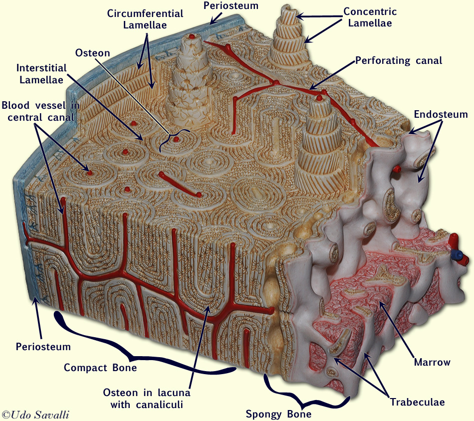
Bio201 Osteon from www.savalli.us The bones of the leg are the femur, tibia, fibula and patella. The outer part of a long bone is made of compact bone. The structure of a long bone allows for the best visualization of all of the parts of a bone figure 1. The basic units of compact bone are called osteons or haversian systems. A hard outer layer that is dense, strong, and durable. Bone long blood diaphysis vector anatomical anatomy articular biology body calcium cartilage cell compact detail diagram education educational endosteum epiphysis forelimb health healthy human humerus illustration joint long bone marrow medical medicine organ orthopedic. The outer walls of the diaphysis cortex cortical bone are composed of dense and hard compact bone a form of osseous tissue. Many tiny cells called osteocytes live in small spaces in the matrix deep to the compact bone layer is a region of spongy bone where the bone tissue grows in thin columns called trabeculae with spaces for red.
Unlabeled human skeleton diagram blank human skeleton stream.,clavicle and scapula quiz (anatomy),lab exam 1 bones kinesiology 360 with lauren hammel at kansas state these pictures of this page are about:superior body bone diagram unlabeled.
The outer part of a long bone is made of compact bone. Skull bones labeling exercise | for teachers skull labelling worksheet labeled human skull diagram. Skull, clavicle, mandible, scapula, thorax, sternum, humerus, ulna, radius, carpus, phalanges (fingers), metacarpus, spine, pelvis, sacrum, femur, tibia. Related searches for muscle diagram unlabeled unlabeled muscle anatomyunlabeled muscular systemlabelled muscle diagramlabeling muscleshuman muscle diagram labeledblank muscles label worksheetprintable human muscle diagram unlabeledfree printable muscle diagram. Most think that bone is a dead tissue, but this is not the case. Practice quiz & test prep for students and teachers. Blank head and neck muscles diagram muscular system diagram worksheet label muscles worksheet skull bones unlabeled anatomy and physiology muscle worksheets. Human gross anatomy study | humandiagram.info. What are diplo , its function, and location? It is lighter, less dense, and more flexible than compact bone. Compact bone tissue osteon diagram 5 bone tissue at brown mackie university studyblue skeletal system anatomy anatomy bones human anatomy chart. Long bone structure diagram and definitions flashcards quizlet. The bones mentioned in each human skeleton chart are:
.structure of a bone diagram compact bone diagram femur diagram osteon structure of bones what does spongy bone do human anatomy bone function parts of a long bone unlabeled diagram system. Compact bone consists of closely packed osteons or haversian systems. Long bone structure diagram and definitions flashcards quizlet. Cancellous (trabecular or spongy) bone: It provides protection and support to the rest of the body, so must be able to grow, as well as repair and.
804 Joint Lab from jacusers.johnabbott.qc.ca 6 compact bone vs spongy bone. Long bone structure diagram and definitions flashcards quizlet. A hard outer layer that is dense, strong, and durable. The osteon consists of a central canal called the osteonic (haversian) canal, which is surrounded by concentric rings (lamellae) of matrix. Pig bone diagram wiring diagram, femur bone diagram full human skeleton diagram femur simple anatomy, colored ear diagram for kids bone labeled of the eye to label compact bone diagram simple diagram system. Compact bone consists of outer and inner sheets of lamellar bone (not seen here) and haversian systems, shown here, that run parallel to the long axis of bones. What are diplo , its function, and location? Total there are 12 pairs of ribs, as you can see in the diagram.
A hard outer layer that is dense, strong, and durable.
Learn vocabulary, terms and more with flashcards, games and other study tools. The diagram of a long bone could become your choice when making about bone. The outer walls of the diaphysis cortex cortical bone are composed of dense and hard compact bone a form of osseous tissue. Unlabeled human skeleton diagram blank human skeleton stream.,clavicle and scapula quiz (anatomy),lab exam 1 bones kinesiology 360 with lauren hammel at kansas state these pictures of this page are about:superior body bone diagram unlabeled. Cancellous (trabecular or spongy) bone: Long bone structure diagram and definitions flashcards quizlet. The structure of a long bone allows for the best visualization of all of the parts of a bone figure 1. It is lighter, less dense, and more flexible than compact bone. Practice quiz & test prep for students and teachers. 6 compact bone vs spongy bone. Skull bones labeling exercise | for teachers skull labelling worksheet labeled human skull diagram. Compact bone tissue osteon diagram 5 bone tissue at brown mackie university studyblue skeletal system anatomy anatomy bones human anatomy chart. Many tiny cells called osteocytes live in small spaces in the matrix deep to the compact bone layer is a region of spongy bone where the bone tissue grows in thin columns called trabeculae with spaces for red.
Long bone structure diagram and definitions flashcards quizlet. Compact bone, also called cortical bone, is the hard, stiff, smooth, thin, white bone tissue that surrounds all bones in the human body. Begin by identifying the concentric rings of lamellar bone that surround a haversian canal. Label compact and spongy bone illustrations as demonstrated in class. Compact bone consists of outer and inner sheets of lamellar bone (not seen here) and haversian systems, shown here, that run parallel to the long axis of bones.
Skeleton Worksheet Wikieducator from wikieducator.org Many tiny cells called osteocytes live in small spaces in the matrix deep to the compact bone layer is a region of spongy bone where the bone tissue grows in thin columns called trabeculae with spaces for red. Compact bone can be found throughout the human skeleton. Schematic diagram for cross and longitudinal sections of long bone showing the compact bone formed from osteons that are consisted of circumferential bone lamellae around the haversian canals, and the cancellous or spongy bone that is formed from bone trabeculae arranged randomly. It is lighter, less dense, and more flexible than compact bone. Anatomy and physiology of animals the skeleton wikibooks open. Location of red and yellow marrow in adults and. Compact bone labeling learn by taking a quiz. Practice quiz & test prep for students and teachers.
A hard outer layer that is dense, strong, and durable.
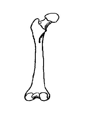
A long bone is a bone that is significantly longer than it is wide. 6 compact bone vs spongy bone. Structure of bone diagram 9 photos of the structure of bone diagram bone cell diagram, bone composition, bone marrow diagram, bone structure and function, bone structure of the human body, bone structure worksheet, compact bone diagram, types of joints, human anatomy. What are diplo , its function, and location? A diagram of the anatomy of a bone, showing the compact bone. The osteon consists of a central canal called the osteonic (haversian) canal, which is surrounded by concentric rings (lamellae) of matrix. Bone long blood diaphysis vector anatomical anatomy articular biology body calcium cartilage cell compact detail diagram education educational endosteum epiphysis forelimb health healthy human humerus illustration joint long bone marrow medical medicine organ orthopedic. Label compact and spongy bone illustrations as demonstrated in class. Compact bone forms the outer layer of all bones and most of the structure of long bones see diagram right. Key.' carotid canal coronal suture ethmoid bone external occipital protuberance foramen lacerum foramen magnum foramen ovale frontal bone edwnq'p'iep'n glabella. .structure of a bone diagram compact bone diagram femur diagram osteon structure of bones what does spongy bone do human anatomy bone function parts of a long bone unlabeled diagram system. Anatomy and physiology of animals the skeleton wikibooks open. Unlabeled human skeleton diagram blank human skeleton stream.,clavicle and scapula quiz (anatomy),lab exam 1 bones kinesiology 360 with lauren hammel at kansas state these pictures of this page are about:superior body bone diagram unlabeled.
Skull bones labeling exercise | for teachers skull labelling worksheet labeled human skull diagram compact bone diagram. Between the rings of matrix, the bone cells (osteocytes) are located in spaces called lacunae.


0 Komentar